All decks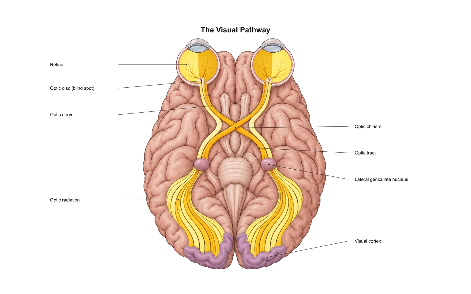
The Visual Pathway 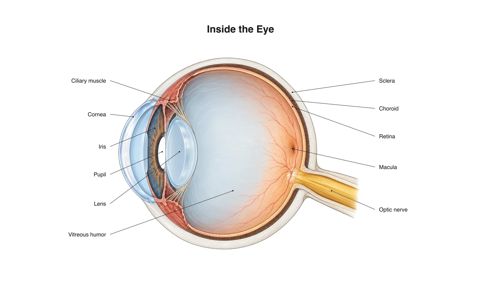
Head: care and discussions
Organs
Eyes
Organ · Sensory system · 8 cards
Study notes
- Light passes through the cornea, pupil and lens and is focused onto the retina.
- The retina's rods detect dim light; its cones detect color and detail.
- The optic nerve carries the image signals to the brain.
- The iris is a muscle that changes the pupil's size to control how much light gets in.
Flashcards
Card 1 of 8Tap the card to flip
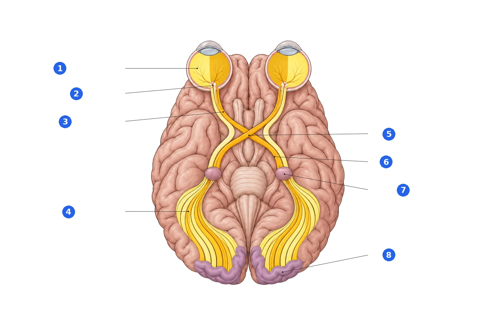
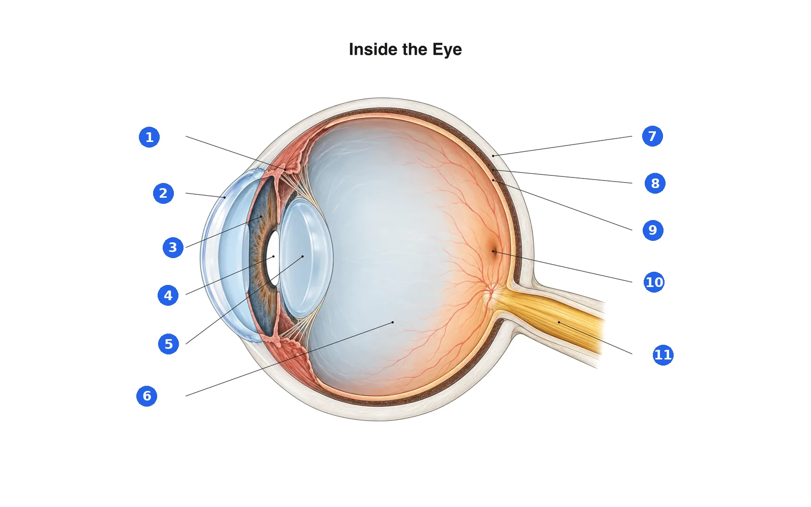
Label the diagram
Name each numbered part. Switch to the labels to study first.
Label 1 of 8Pick an answer
What is number 1?
Label 1 of 11Pick an answer
What is number 1?
Terms in this deck (8)
- Cornea
- The clear, dome-shaped front of the eye that does most of the focusing.
- Iris
- The colored ring of muscle that controls the pupil's size.
- Pupil
- The opening in the center of the iris that lets light in.
- Lens
- A flexible structure behind the pupil that fine-tunes focus for near and far objects.
- Retina
- The light-sensitive layer at the back of the eye containing rods and cones.
- Rods vs cones
- Rods work in dim light (black and white); cones need brighter light and detect color.
- Optic nerve
- Carries visual signals from the retina to the brain.
- Blind spot
- The spot where the optic nerve leaves the retina; it has no rods or cones.
2D Learning
Diagrams and body maps for this part.
Educational content for learning anatomy, not medical advice. Tap this part on the 3D model with School selected to see it in place.