2D Learning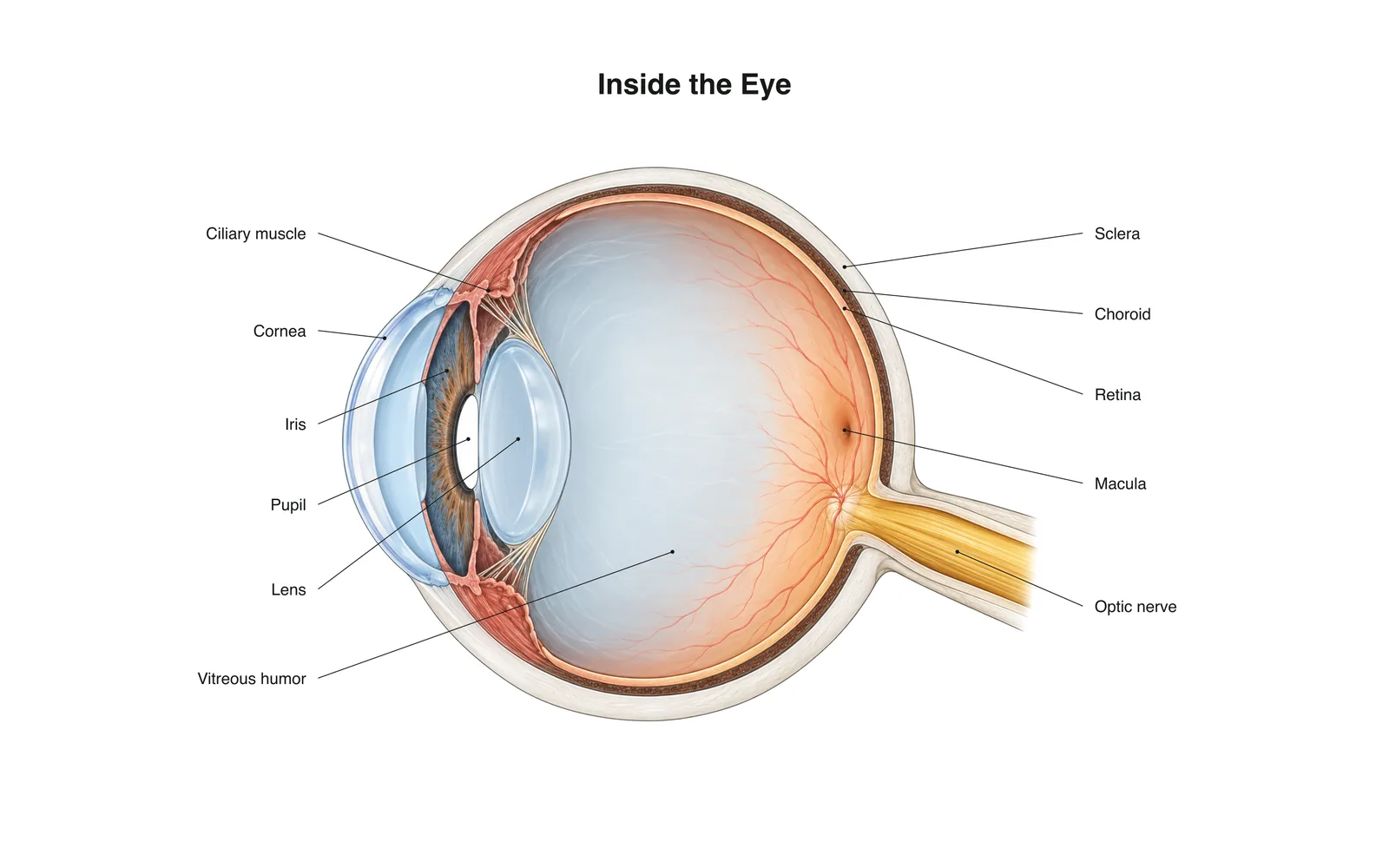
2D Learning
Inside the Eye
How it works · Body maps · 11 cards
Study notes
- Light passes through the cornea and lens, which focus it onto the retina.
- The retina's rods and cones turn light into nerve signals sent along the optic nerve.
- The image on the retina is upside down; the brain flips it.
Flashcards
Card 1 of 11Tap the card to flip
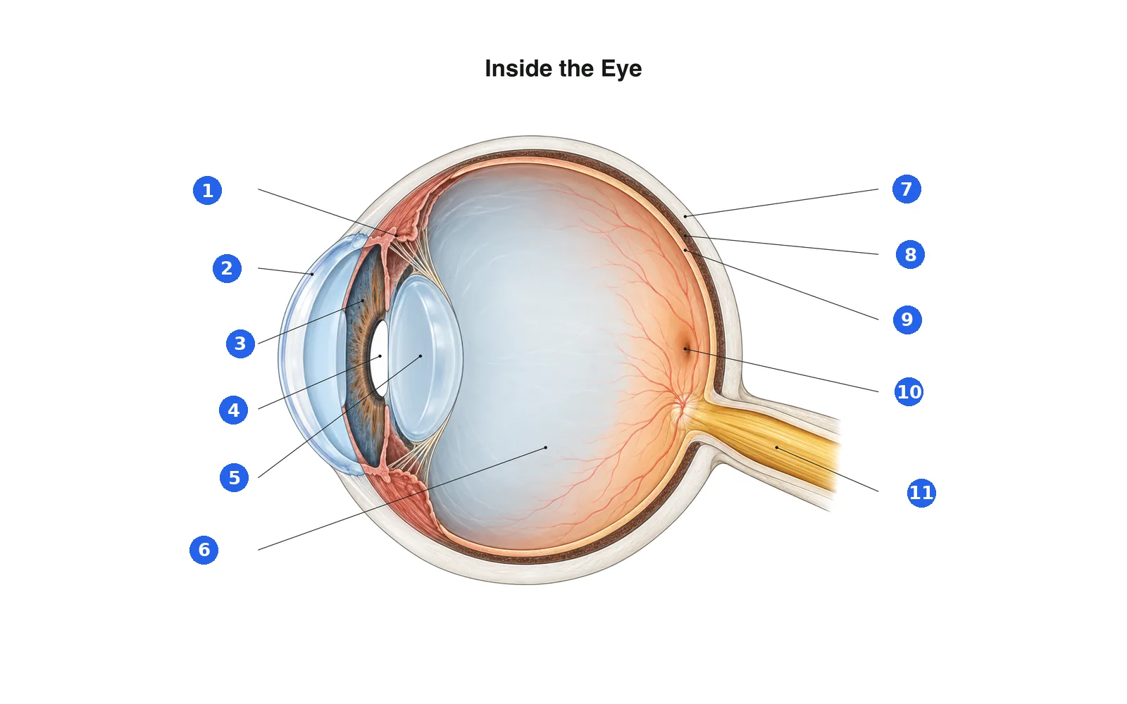
Label the diagram
Name each numbered part. Switch to the labels to study first.
Label 1 of 11Pick an answer
What is number 1?
Terms in this deck (11)
- Cornea
- The clear dome at the front of the eye that does most of the focusing.
- Iris
- The colored ring that widens or narrows the pupil.
- Pupil
- The opening in the iris that lets light in.
- Lens
- Changes shape to fine-focus on near or far objects.
- Ciliary muscle
- Ring of muscle that changes the lens's shape for focusing.
- Sclera
- The tough white outer layer of the eye.
- Choroid
- The blood-rich layer between the sclera and retina that feeds the retina.
- Retina
- The light-sensing layer at the back of the eye.
- Macula
- The center of the retina, packed with cones for sharp, detailed vision.
- Optic nerve
- Carries signals from the retina to the brain.
- Vitreous humor
- The clear gel that fills the eyeball and keeps its shape.
See it on the 3D body
Open the 3D model on a part, with its School notes and flashcards.
Educational content for learning anatomy, not medical advice.