Apprentissage 2D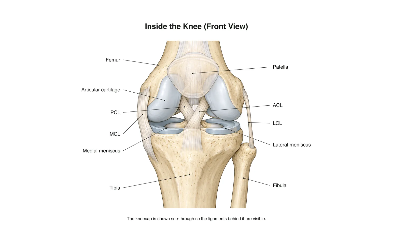
Apprentissage 2D
Inside the Knee
How it works · Body maps · 11 cartes
Notes d'étude
- The knee is the body's largest joint: a hinge between the femur and tibia.
- Four main ligaments hold it together: two crossing inside (ACL, PCL) and two at the sides (MCL, LCL).
- The menisci are C-shaped cartilage pads that cushion and steady the joint.
Cartes mémoire
Carte 1 sur 11Touchez la carte pour la retourner
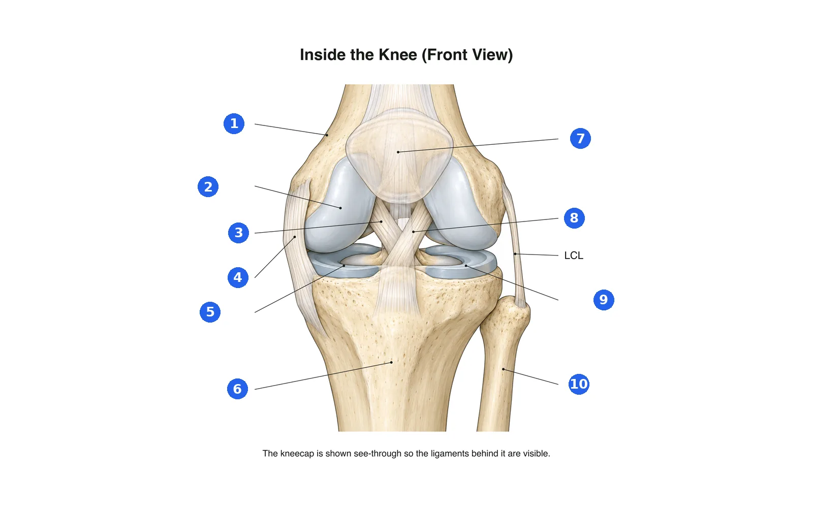
Légende le schéma
Nomme chaque partie numérotée. Passe aux légendes pour étudier d'abord.
Légende 1 sur 10Choisissez une réponse
Quel est le numéro 1 ?
Termes de ce paquet (11)
- Femur
- The thigh bone, forming the top of the knee.
- Tibia
- The shin bone, forming the bottom of the knee.
- Fibula
- The thin outer leg bone; not part of the knee joint itself.
- Patella
- The kneecap, sliding in a groove at the front of the femur.
- Articular cartilage
- Smooth covering on the bone ends that lets them glide.
- Medial meniscus
- The inner C-shaped cartilage pad between femur and tibia.
- Lateral meniscus
- The outer C-shaped cartilage pad between femur and tibia.
- ACL
- Anterior cruciate ligament: stops the tibia sliding forward; often torn in sport.
- PCL
- Posterior cruciate ligament: stops the tibia sliding backward.
- MCL
- Medial collateral ligament on the inner side of the knee.
- LCL
- Lateral collateral ligament on the outer side of the knee.
Le voir sur le corps 3D
Ouvrez le modèle 3D sur une partie, avec ses notes et fiches d'École.
Contenu éducatif pour apprendre l'anatomie, pas un avis médical.