Apprentissage 2D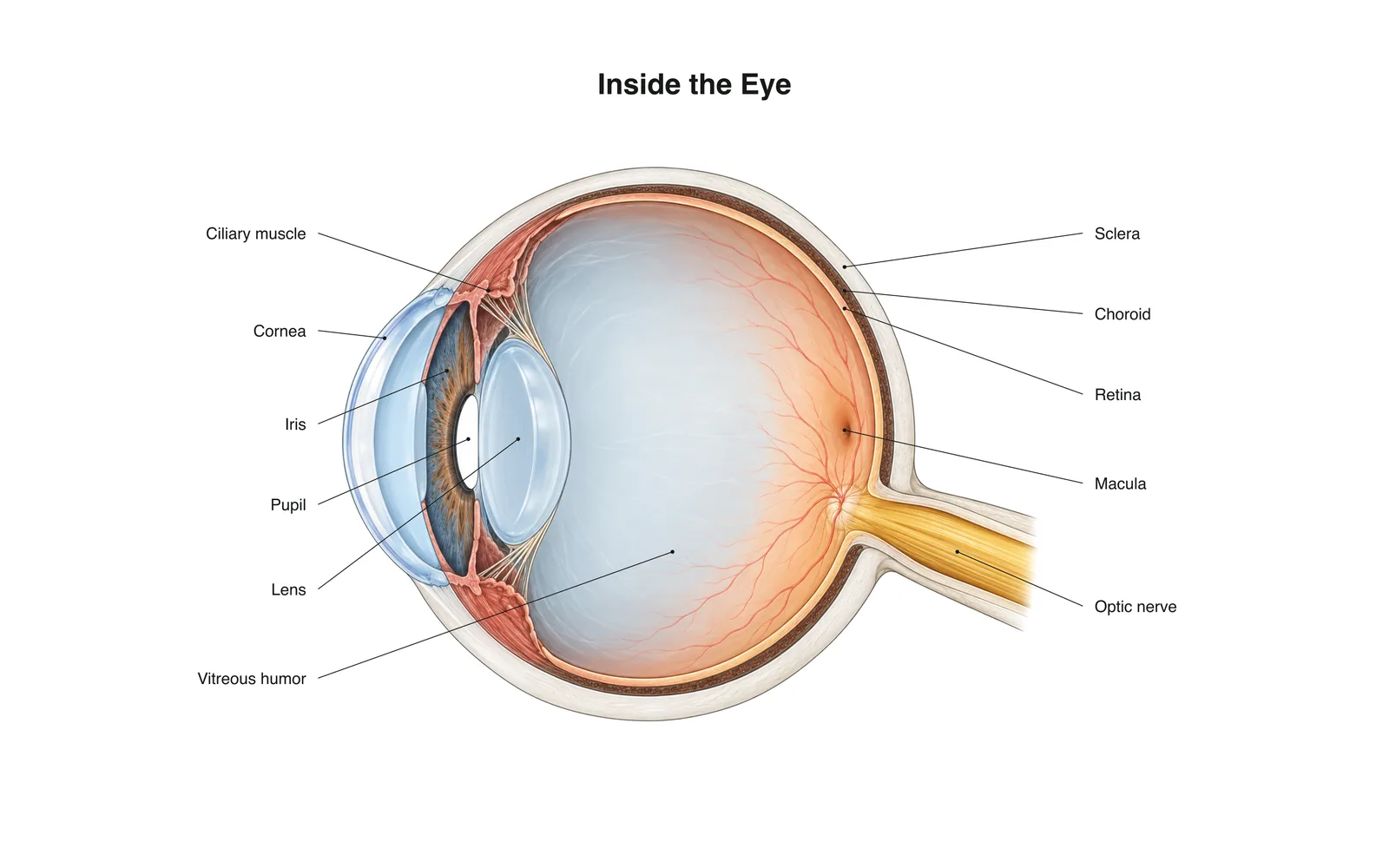
Apprentissage 2D
Inside the Eye
How it works · Body maps · 11 cartes
Notes d'étude
- Light passes through the cornea and lens, which focus it onto the retina.
- The retina's rods and cones turn light into nerve signals sent along the optic nerve.
- The image on the retina is upside down; the brain flips it.
Cartes mémoire
Carte 1 sur 11Touchez la carte pour la retourner
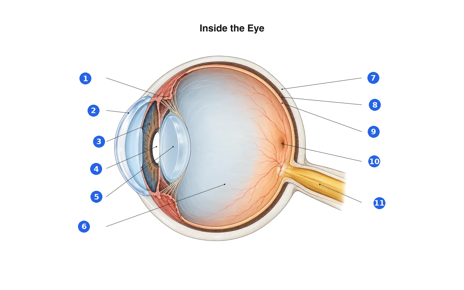
Légende le schéma
Nomme chaque partie numérotée. Passe aux légendes pour étudier d'abord.
Légende 1 sur 11Choisissez une réponse
Quel est le numéro 1 ?
Termes de ce paquet (11)
- Cornea
- The clear dome at the front of the eye that does most of the focusing.
- Iris
- The colored ring that widens or narrows the pupil.
- Pupil
- The opening in the iris that lets light in.
- Lens
- Changes shape to fine-focus on near or far objects.
- Ciliary muscle
- Ring of muscle that changes the lens's shape for focusing.
- Sclera
- The tough white outer layer of the eye.
- Choroid
- The blood-rich layer between the sclera and retina that feeds the retina.
- Retina
- The light-sensing layer at the back of the eye.
- Macula
- The center of the retina, packed with cones for sharp, detailed vision.
- Optic nerve
- Carries signals from the retina to the brain.
- Vitreous humor
- The clear gel that fills the eyeball and keeps its shape.
Le voir sur le corps 3D
Ouvrez le modèle 3D sur une partie, avec ses notes et fiches d'École.
Contenu éducatif pour apprendre l'anatomie, pas un avis médical.