Aprendizaje 2D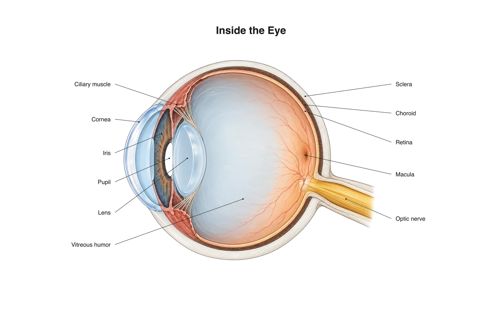
Aprendizaje 2D
Inside the Eye
How it works · Body maps · 11 tarjetas
Notas de estudio
- Light passes through the cornea and lens, which focus it onto the retina.
- The retina's rods and cones turn light into nerve signals sent along the optic nerve.
- The image on the retina is upside down; the brain flips it.
Tarjetas
Tarjeta 1 de 11Toca la tarjeta para darle la vuelta
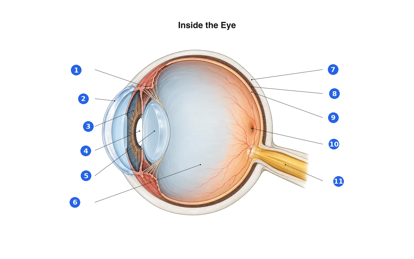
Etiqueta el diagrama
Nombra cada parte numerada. Cambia a las etiquetas para estudiar primero.
Etiqueta 1 de 11Elige una respuesta
¿Qué es el número 1?
Términos de este mazo (11)
- Cornea
- The clear dome at the front of the eye that does most of the focusing.
- Iris
- The colored ring that widens or narrows the pupil.
- Pupil
- The opening in the iris that lets light in.
- Lens
- Changes shape to fine-focus on near or far objects.
- Ciliary muscle
- Ring of muscle that changes the lens's shape for focusing.
- Sclera
- The tough white outer layer of the eye.
- Choroid
- The blood-rich layer between the sclera and retina that feeds the retina.
- Retina
- The light-sensing layer at the back of the eye.
- Macula
- The center of the retina, packed with cones for sharp, detailed vision.
- Optic nerve
- Carries signals from the retina to the brain.
- Vitreous humor
- The clear gel that fills the eyeball and keeps its shape.
Verlo en el cuerpo 3D
Abre el modelo 3D en una parte, con sus notas y tarjetas de Escuela.
Contenido educativo para aprender anatomía, no consejo médico.