Kujifunza kwa 2D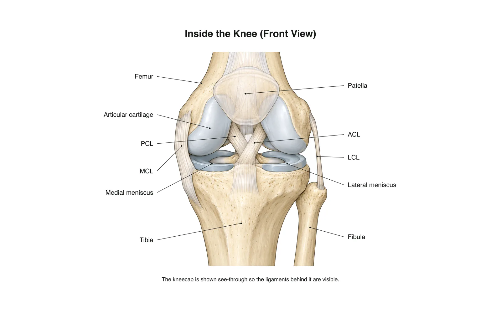
Kujifunza kwa 2D
Inside the Knee
How it works · Body maps · Kadi 11
Maelezo ya kusoma
- The knee is the body's largest joint: a hinge between the femur and tibia.
- Four main ligaments hold it together: two crossing inside (ACL, PCL) and two at the sides (MCL, LCL).
- The menisci are C-shaped cartilage pads that cushion and steady the joint.
Kadi za kujifunzia
Kadi 1 kati ya 11Gusa kadi kuigeuza
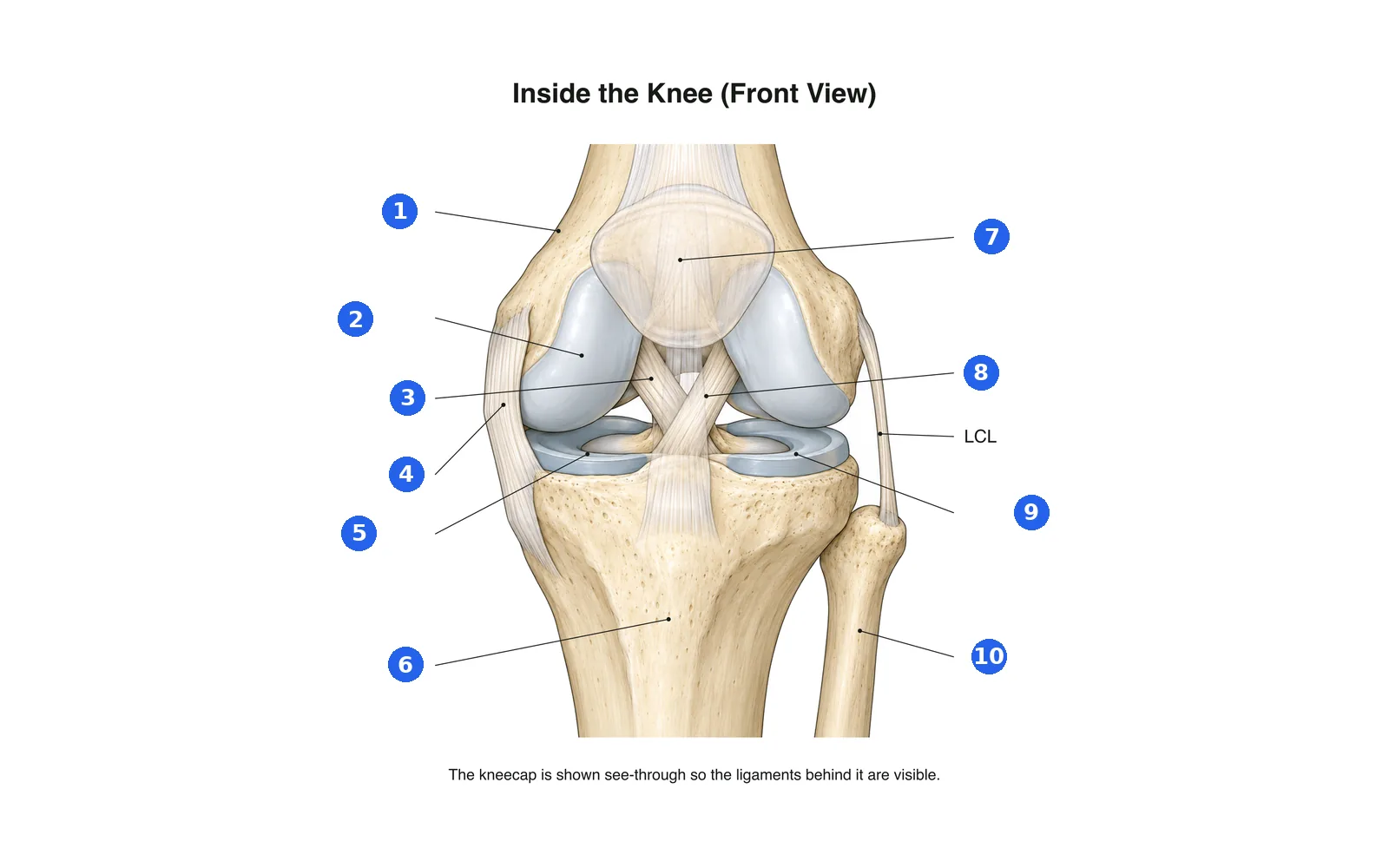
Taja sehemu za mchoro
Taja kila sehemu yenye namba. Badilisha kwenda kwenye majina ili usome kwanza.
Jina 1 kati ya 10Chagua jibu
Namba 1 ni nini?
Maneno katika seti hii (11)
- Femur
- The thigh bone, forming the top of the knee.
- Tibia
- The shin bone, forming the bottom of the knee.
- Fibula
- The thin outer leg bone; not part of the knee joint itself.
- Patella
- The kneecap, sliding in a groove at the front of the femur.
- Articular cartilage
- Smooth covering on the bone ends that lets them glide.
- Medial meniscus
- The inner C-shaped cartilage pad between femur and tibia.
- Lateral meniscus
- The outer C-shaped cartilage pad between femur and tibia.
- ACL
- Anterior cruciate ligament: stops the tibia sliding forward; often torn in sport.
- PCL
- Posterior cruciate ligament: stops the tibia sliding backward.
- MCL
- Medial collateral ligament on the inner side of the knee.
- LCL
- Lateral collateral ligament on the outer side of the knee.
Ione kwenye mwili wa 3D
Fungua modeli ya 3D kwenye sehemu, pamoja na maelezo na kadi zake za Shule.
Maudhui ya kielimu ya kujifunza anatomia, si ushauri wa kitabibu.