Kujifunza kwa 2D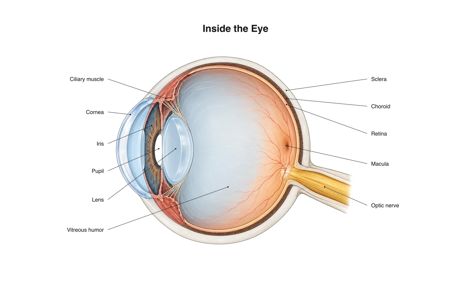
Kujifunza kwa 2D
Inside the Eye
How it works · Body maps · Kadi 11
Maelezo ya kusoma
- Light passes through the cornea and lens, which focus it onto the retina.
- The retina's rods and cones turn light into nerve signals sent along the optic nerve.
- The image on the retina is upside down; the brain flips it.
Kadi za kujifunzia
Kadi 1 kati ya 11Gusa kadi kuigeuza
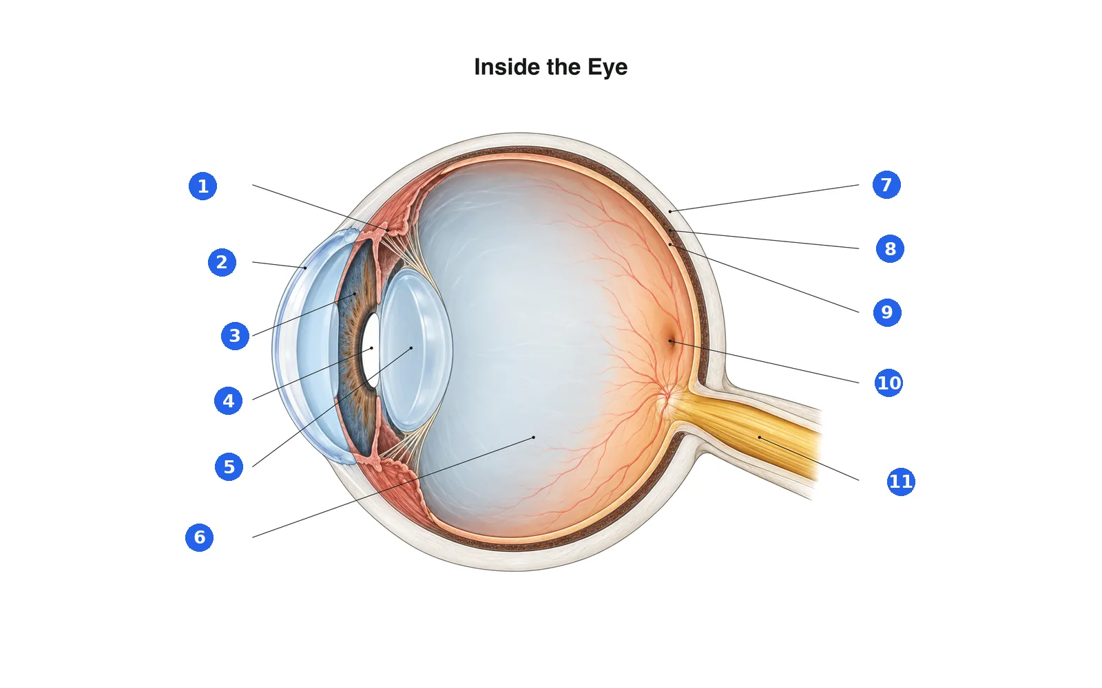
Taja sehemu za mchoro
Taja kila sehemu yenye namba. Badilisha kwenda kwenye majina ili usome kwanza.
Jina 1 kati ya 11Chagua jibu
Namba 1 ni nini?
Maneno katika seti hii (11)
- Cornea
- The clear dome at the front of the eye that does most of the focusing.
- Iris
- The colored ring that widens or narrows the pupil.
- Pupil
- The opening in the iris that lets light in.
- Lens
- Changes shape to fine-focus on near or far objects.
- Ciliary muscle
- Ring of muscle that changes the lens's shape for focusing.
- Sclera
- The tough white outer layer of the eye.
- Choroid
- The blood-rich layer between the sclera and retina that feeds the retina.
- Retina
- The light-sensing layer at the back of the eye.
- Macula
- The center of the retina, packed with cones for sharp, detailed vision.
- Optic nerve
- Carries signals from the retina to the brain.
- Vitreous humor
- The clear gel that fills the eyeball and keeps its shape.
Ione kwenye mwili wa 3D
Fungua modeli ya 3D kwenye sehemu, pamoja na maelezo na kadi zake za Shule.
Maudhui ya kielimu ya kujifunza anatomia, si ushauri wa kitabibu.