Aprendizagem 2D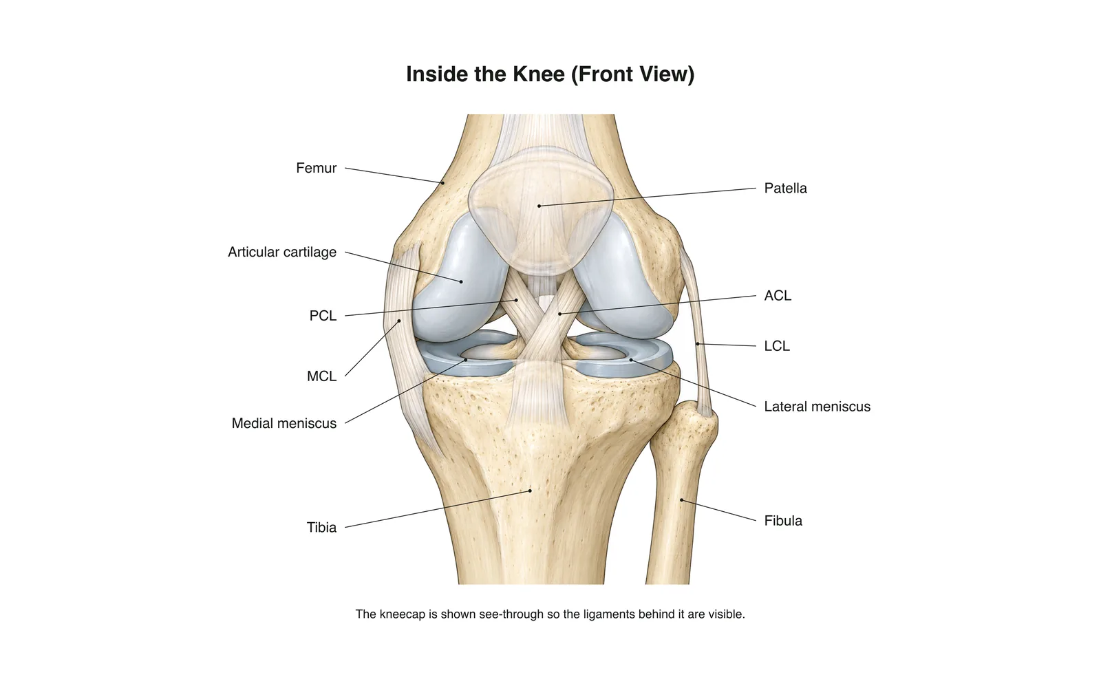
Aprendizagem 2D
Inside the Knee
How it works · Body maps · 11 cartões
Notas de estudo
- The knee is the body's largest joint: a hinge between the femur and tibia.
- Four main ligaments hold it together: two crossing inside (ACL, PCL) and two at the sides (MCL, LCL).
- The menisci are C-shaped cartilage pads that cushion and steady the joint.
Flashcards
Cartão 1 de 11Toque no cartão para virar
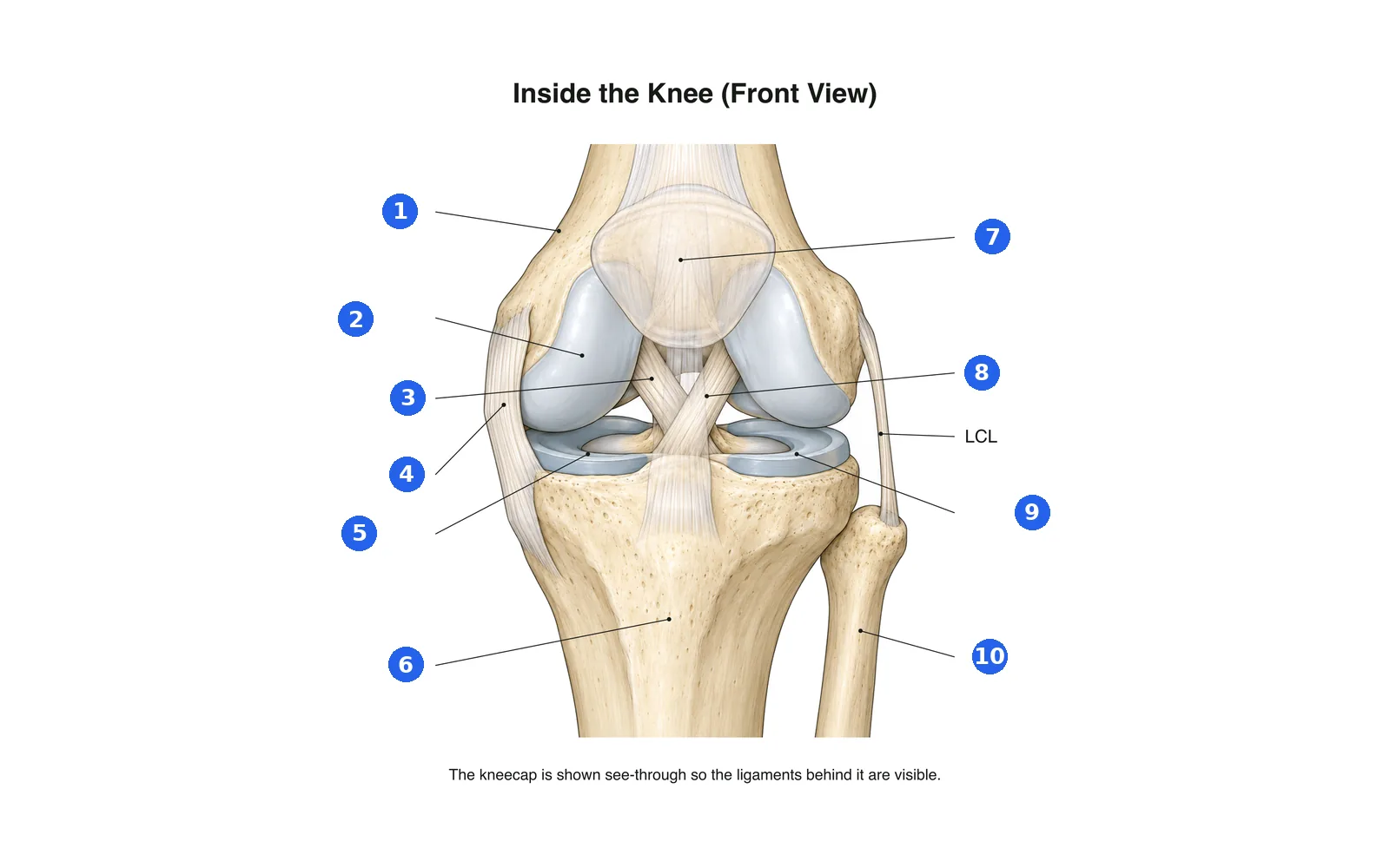
Identifique o diagrama
Nomeie cada parte numerada. Mude para os rótulos para estudar primeiro.
Rótulo 1 de 10Escolha uma resposta
Qual é o número 1?
Termos deste baralho (11)
- Femur
- The thigh bone, forming the top of the knee.
- Tibia
- The shin bone, forming the bottom of the knee.
- Fibula
- The thin outer leg bone; not part of the knee joint itself.
- Patella
- The kneecap, sliding in a groove at the front of the femur.
- Articular cartilage
- Smooth covering on the bone ends that lets them glide.
- Medial meniscus
- The inner C-shaped cartilage pad between femur and tibia.
- Lateral meniscus
- The outer C-shaped cartilage pad between femur and tibia.
- ACL
- Anterior cruciate ligament: stops the tibia sliding forward; often torn in sport.
- PCL
- Posterior cruciate ligament: stops the tibia sliding backward.
- MCL
- Medial collateral ligament on the inner side of the knee.
- LCL
- Lateral collateral ligament on the outer side of the knee.
Ver no corpo 3D
Abra o modelo 3D numa parte, com as notas e cartões da Escola.
Conteúdo educativo para aprender anatomia, não é aconselhamento médico.