2D लर्निंग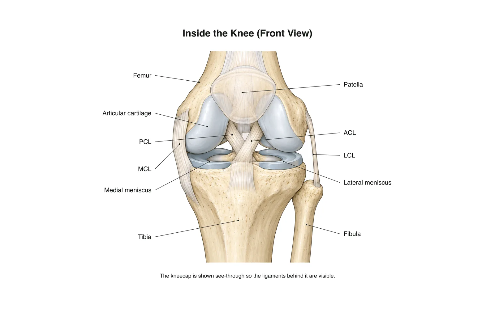
2D लर्निंग
Inside the Knee
How it works · Body maps · 11 कार्ड
अध्ययन नोट्स
- The knee is the body's largest joint: a hinge between the femur and tibia.
- Four main ligaments hold it together: two crossing inside (ACL, PCL) and two at the sides (MCL, LCL).
- The menisci are C-shaped cartilage pads that cushion and steady the joint.
फ़्लैशकार्ड
कार्ड 1 / 11पलटने के लिए कार्ड टैप करें
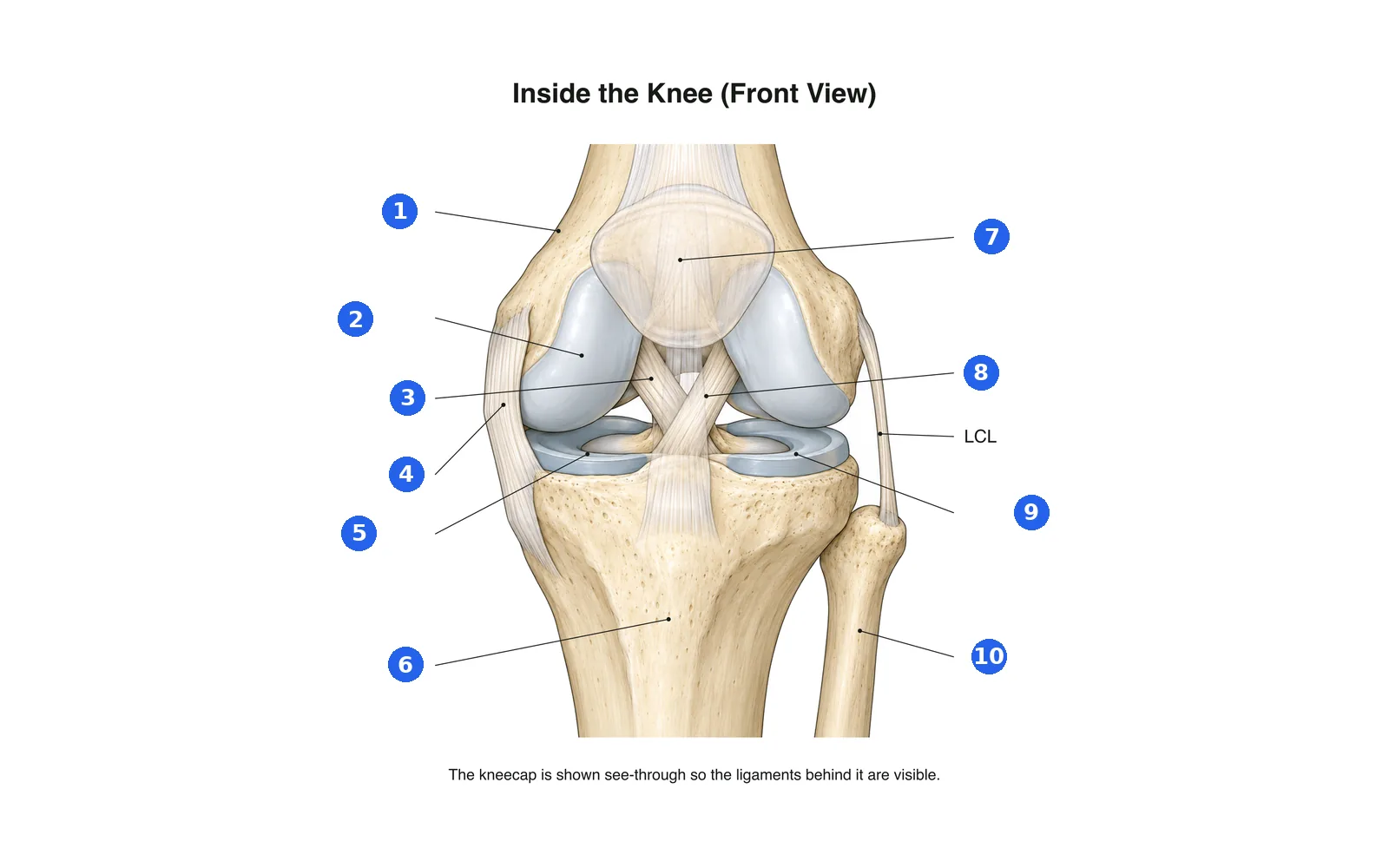
चित्र के भाग पहचानें
हर क्रमांकित भाग का नाम बताएँ। पहले पढ़ने के लिए लेबल देखें।
लेबल 1 / 10एक उत्तर चुनें
संख्या 1 क्या है?
इस डेक के शब्द (11)
- Femur
- The thigh bone, forming the top of the knee.
- Tibia
- The shin bone, forming the bottom of the knee.
- Fibula
- The thin outer leg bone; not part of the knee joint itself.
- Patella
- The kneecap, sliding in a groove at the front of the femur.
- Articular cartilage
- Smooth covering on the bone ends that lets them glide.
- Medial meniscus
- The inner C-shaped cartilage pad between femur and tibia.
- Lateral meniscus
- The outer C-shaped cartilage pad between femur and tibia.
- ACL
- Anterior cruciate ligament: stops the tibia sliding forward; often torn in sport.
- PCL
- Posterior cruciate ligament: stops the tibia sliding backward.
- MCL
- Medial collateral ligament on the inner side of the knee.
- LCL
- Lateral collateral ligament on the outer side of the knee.
इसे 3D शरीर पर देखें
3D मॉडल को किसी हिस्से पर खोलें, उसके स्कूल नोट्स और फ्लैशकार्ड के साथ।
शरीर रचना सीखने के लिए शैक्षिक सामग्री, चिकित्सा सलाह नहीं।