2D लर्निंग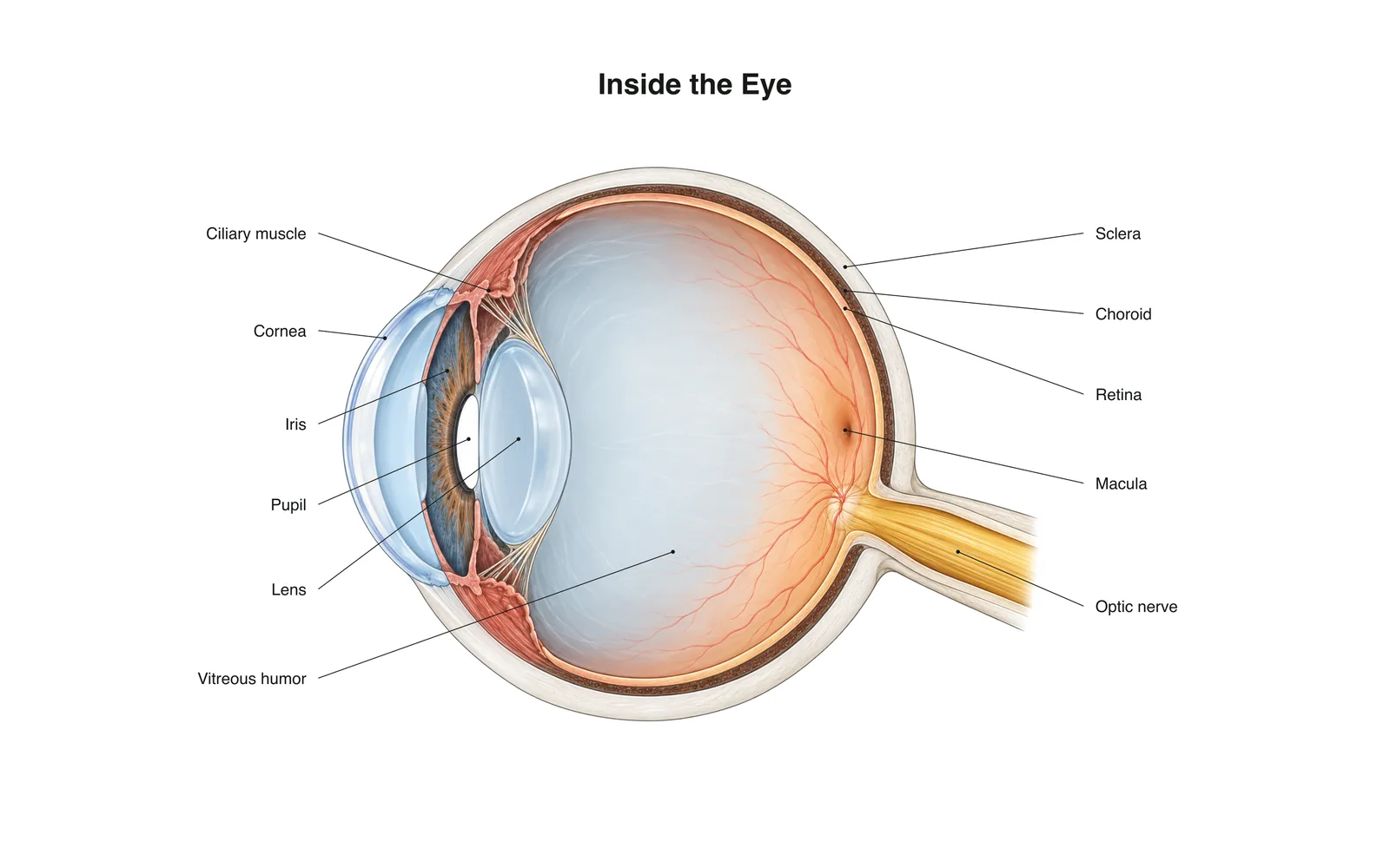
2D लर्निंग
Inside the Eye
How it works · Body maps · 11 कार्ड
अध्ययन नोट्स
- Light passes through the cornea and lens, which focus it onto the retina.
- The retina's rods and cones turn light into nerve signals sent along the optic nerve.
- The image on the retina is upside down; the brain flips it.
फ़्लैशकार्ड
कार्ड 1 / 11पलटने के लिए कार्ड टैप करें
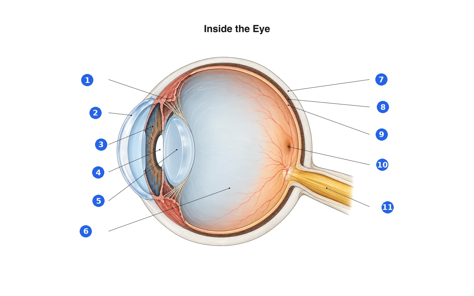
चित्र के भाग पहचानें
हर क्रमांकित भाग का नाम बताएँ। पहले पढ़ने के लिए लेबल देखें।
लेबल 1 / 11एक उत्तर चुनें
संख्या 1 क्या है?
इस डेक के शब्द (11)
- Cornea
- The clear dome at the front of the eye that does most of the focusing.
- Iris
- The colored ring that widens or narrows the pupil.
- Pupil
- The opening in the iris that lets light in.
- Lens
- Changes shape to fine-focus on near or far objects.
- Ciliary muscle
- Ring of muscle that changes the lens's shape for focusing.
- Sclera
- The tough white outer layer of the eye.
- Choroid
- The blood-rich layer between the sclera and retina that feeds the retina.
- Retina
- The light-sensing layer at the back of the eye.
- Macula
- The center of the retina, packed with cones for sharp, detailed vision.
- Optic nerve
- Carries signals from the retina to the brain.
- Vitreous humor
- The clear gel that fills the eyeball and keeps its shape.
इसे 3D शरीर पर देखें
3D मॉडल को किसी हिस्से पर खोलें, उसके स्कूल नोट्स और फ्लैशकार्ड के साथ।
शरीर रचना सीखने के लिए शैक्षिक सामग्री, चिकित्सा सलाह नहीं।