Aprendizaje 2D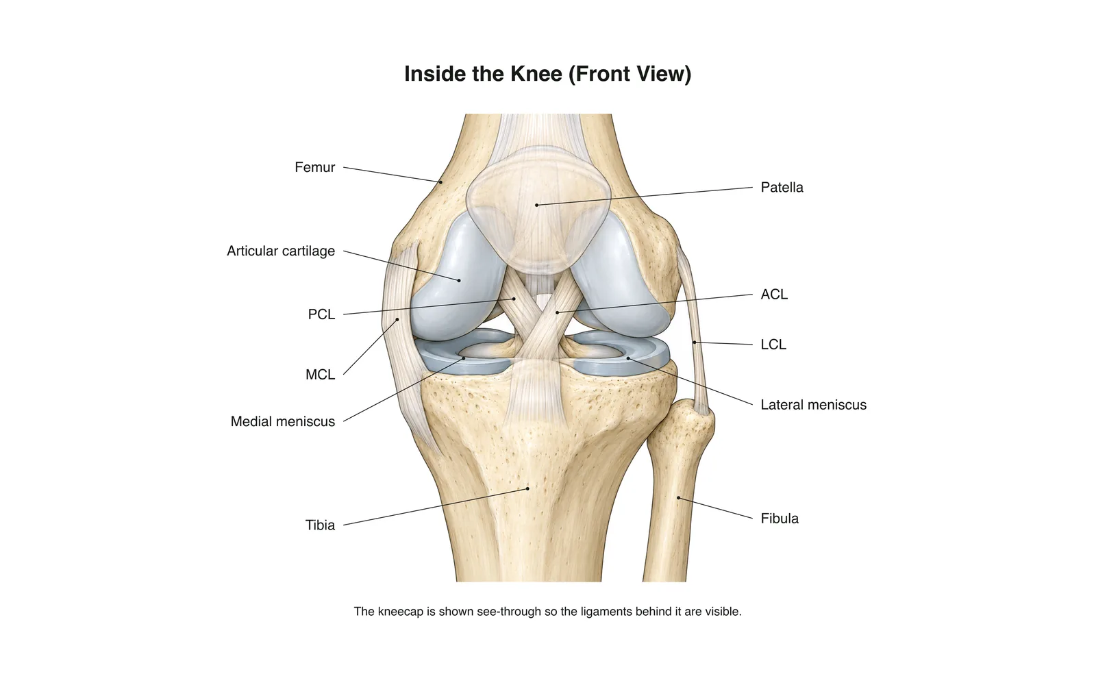
Aprendizaje 2D
Inside the Knee
How it works · Body maps · 11 tarjetas
Notas de estudio
- The knee is the body's largest joint: a hinge between the femur and tibia.
- Four main ligaments hold it together: two crossing inside (ACL, PCL) and two at the sides (MCL, LCL).
- The menisci are C-shaped cartilage pads that cushion and steady the joint.
Tarjetas
Tarjeta 1 de 11Toca la tarjeta para darle la vuelta
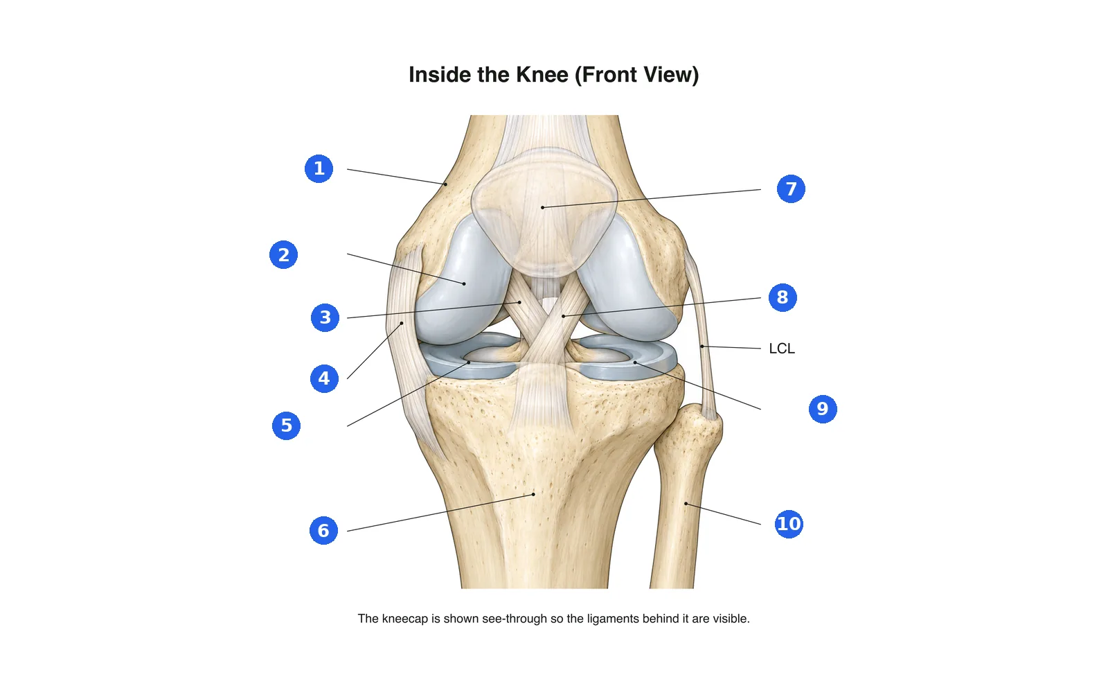
Etiqueta el diagrama
Nombra cada parte numerada. Cambia a las etiquetas para estudiar primero.
Etiqueta 1 de 10Elige una respuesta
¿Qué es el número 1?
Términos de este mazo (11)
- Femur
- The thigh bone, forming the top of the knee.
- Tibia
- The shin bone, forming the bottom of the knee.
- Fibula
- The thin outer leg bone; not part of the knee joint itself.
- Patella
- The kneecap, sliding in a groove at the front of the femur.
- Articular cartilage
- Smooth covering on the bone ends that lets them glide.
- Medial meniscus
- The inner C-shaped cartilage pad between femur and tibia.
- Lateral meniscus
- The outer C-shaped cartilage pad between femur and tibia.
- ACL
- Anterior cruciate ligament: stops the tibia sliding forward; often torn in sport.
- PCL
- Posterior cruciate ligament: stops the tibia sliding backward.
- MCL
- Medial collateral ligament on the inner side of the knee.
- LCL
- Lateral collateral ligament on the outer side of the knee.
Verlo en el cuerpo 3D
Abre el modelo 3D en una parte, con sus notas y tarjetas de Escuela.
Contenido educativo para aprender anatomía, no consejo médico.