التعلّم ثنائي الأبعاد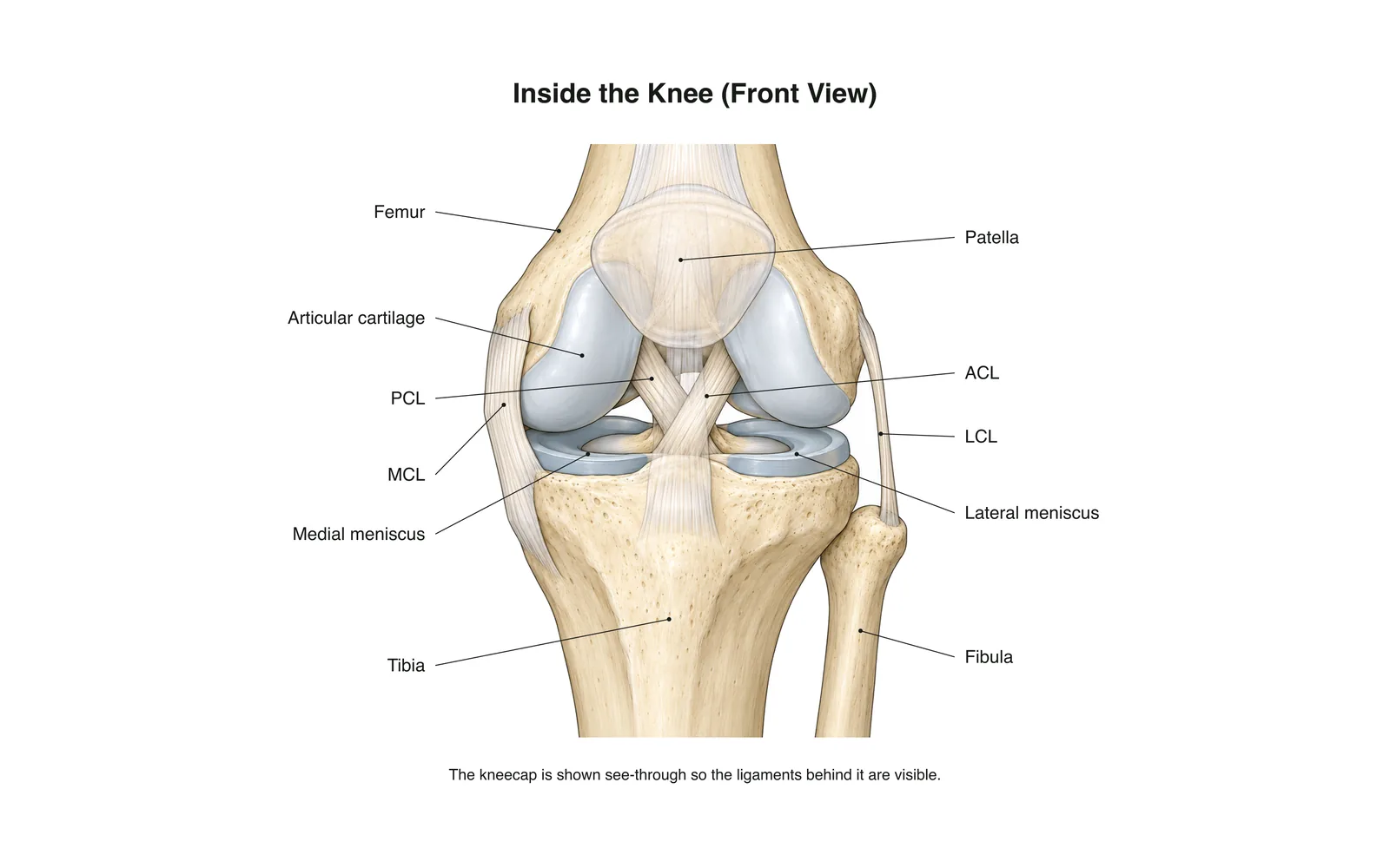
التعلّم ثنائي الأبعاد
Inside the Knee
How it works · Body maps · 11 بطاقة
ملاحظات الدراسة
- The knee is the body's largest joint: a hinge between the femur and tibia.
- Four main ligaments hold it together: two crossing inside (ACL, PCL) and two at the sides (MCL, LCL).
- The menisci are C-shaped cartilage pads that cushion and steady the joint.
البطاقات التعليمية
البطاقة 1 من 11المس البطاقة لقلبها
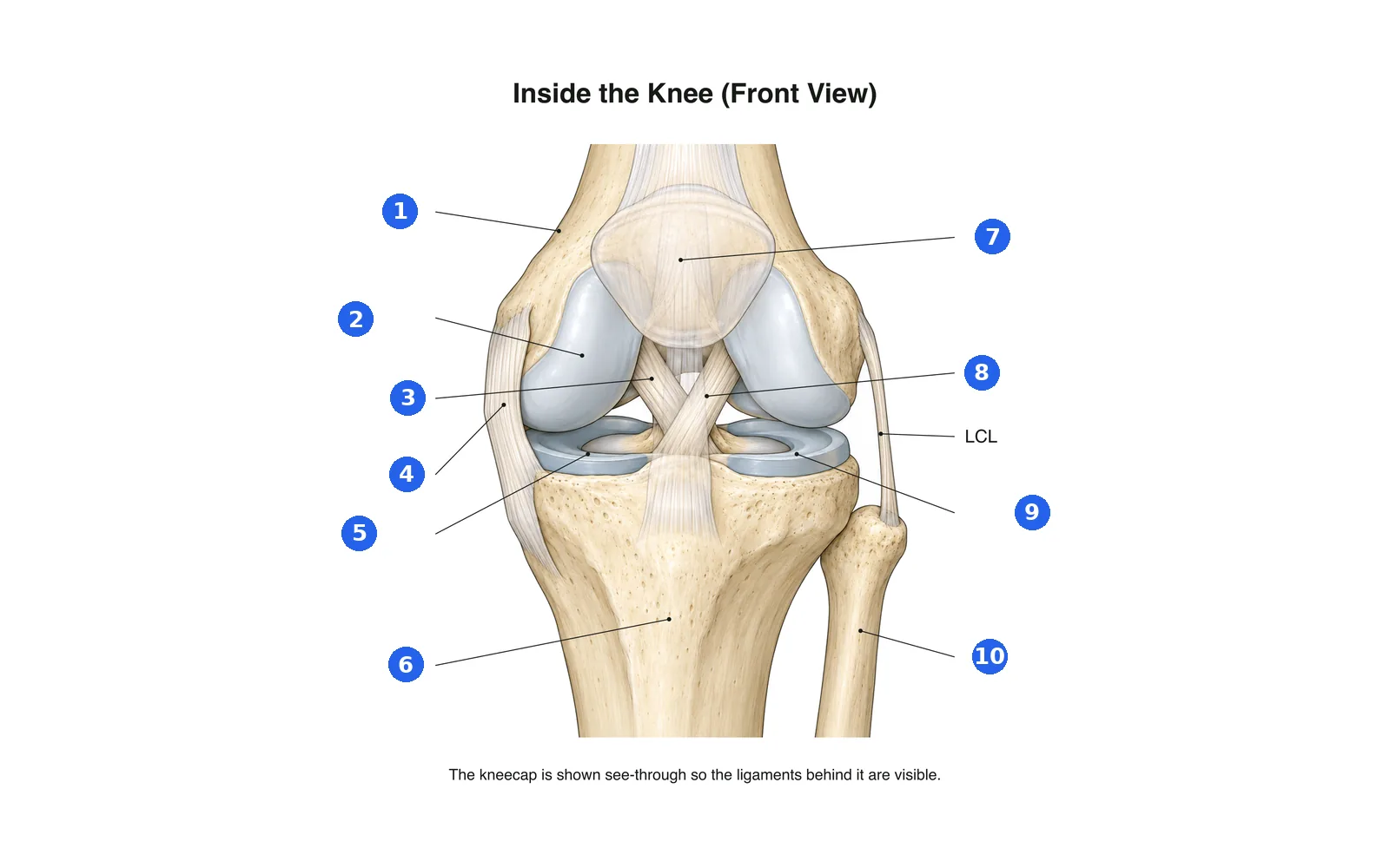
سمِّ أجزاء الرسم
سمِّ كل جزء مرقّم. انتقل إلى التسميات لتدرس أولاً.
التسمية 1 من 10اختر إجابة
ما هو الرقم 1؟
مصطلحات هذه المجموعة (11)
- Femur
- The thigh bone, forming the top of the knee.
- Tibia
- The shin bone, forming the bottom of the knee.
- Fibula
- The thin outer leg bone; not part of the knee joint itself.
- Patella
- The kneecap, sliding in a groove at the front of the femur.
- Articular cartilage
- Smooth covering on the bone ends that lets them glide.
- Medial meniscus
- The inner C-shaped cartilage pad between femur and tibia.
- Lateral meniscus
- The outer C-shaped cartilage pad between femur and tibia.
- ACL
- Anterior cruciate ligament: stops the tibia sliding forward; often torn in sport.
- PCL
- Posterior cruciate ligament: stops the tibia sliding backward.
- MCL
- Medial collateral ligament on the inner side of the knee.
- LCL
- Lateral collateral ligament on the outer side of the knee.
شاهده على الجسم ثلاثي الأبعاد
افتح النموذج ثلاثي الأبعاد على جزء، مع ملاحظاته وبطاقاته في المدرسة.
محتوى تعليمي لتعلم التشريح، وليس نصيحة طبية.