التعلّم ثنائي الأبعاد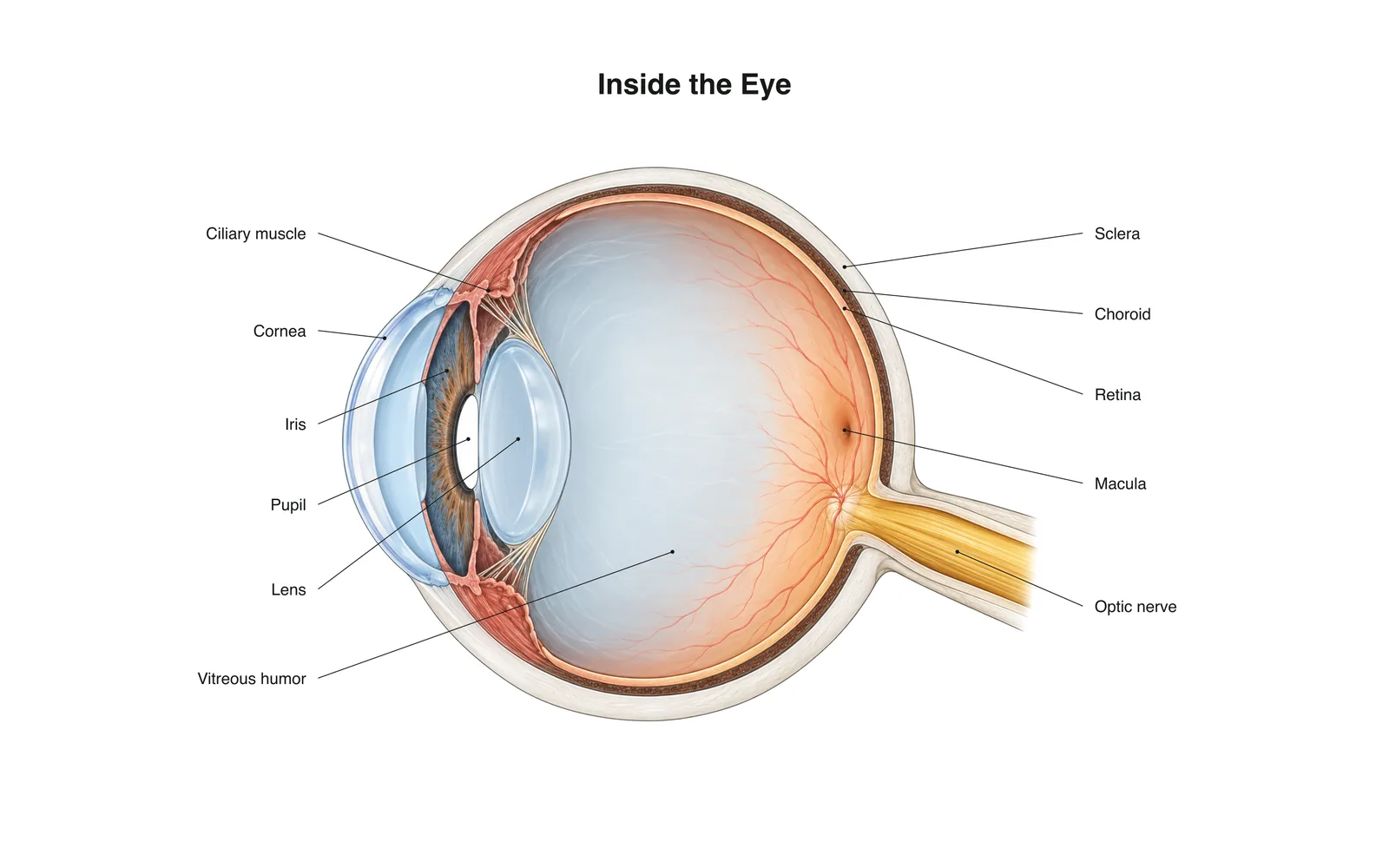
التعلّم ثنائي الأبعاد
Inside the Eye
How it works · Body maps · 11 بطاقة
ملاحظات الدراسة
- Light passes through the cornea and lens, which focus it onto the retina.
- The retina's rods and cones turn light into nerve signals sent along the optic nerve.
- The image on the retina is upside down; the brain flips it.
البطاقات التعليمية
البطاقة 1 من 11المس البطاقة لقلبها
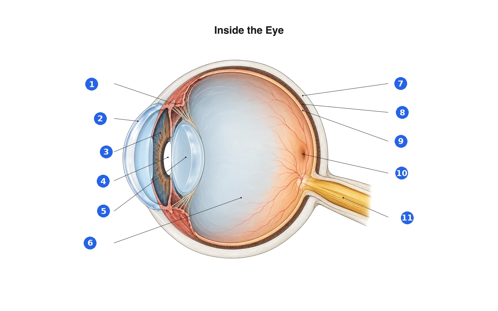
سمِّ أجزاء الرسم
سمِّ كل جزء مرقّم. انتقل إلى التسميات لتدرس أولاً.
التسمية 1 من 11اختر إجابة
ما هو الرقم 1؟
مصطلحات هذه المجموعة (11)
- Cornea
- The clear dome at the front of the eye that does most of the focusing.
- Iris
- The colored ring that widens or narrows the pupil.
- Pupil
- The opening in the iris that lets light in.
- Lens
- Changes shape to fine-focus on near or far objects.
- Ciliary muscle
- Ring of muscle that changes the lens's shape for focusing.
- Sclera
- The tough white outer layer of the eye.
- Choroid
- The blood-rich layer between the sclera and retina that feeds the retina.
- Retina
- The light-sensing layer at the back of the eye.
- Macula
- The center of the retina, packed with cones for sharp, detailed vision.
- Optic nerve
- Carries signals from the retina to the brain.
- Vitreous humor
- The clear gel that fills the eyeball and keeps its shape.
شاهده على الجسم ثلاثي الأبعاد
افتح النموذج ثلاثي الأبعاد على جزء، مع ملاحظاته وبطاقاته في المدرسة.
محتوى تعليمي لتعلم التشريح، وليس نصيحة طبية.