2D ትምህርት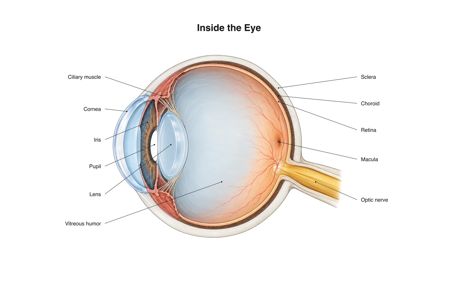
2D ትምህርት
Inside the Eye
How it works · Body maps · 11 ካርዶች
የጥናት ማስታወሻዎች
- Light passes through the cornea and lens, which focus it onto the retina.
- The retina's rods and cones turn light into nerve signals sent along the optic nerve.
- The image on the retina is upside down; the brain flips it.
የማስታወሻ ካርዶች
ካርድ 1 ከ11ለመገልበጥ ካርዱን ይንኩ
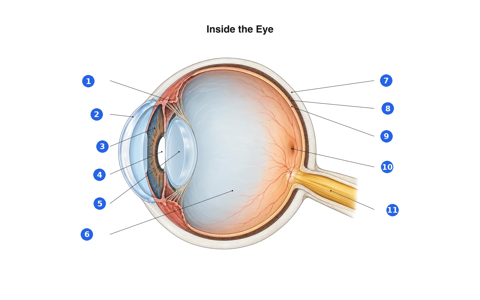
የሥዕሉን ክፍሎች ሰይም
ቁጥር ያለውን እያንዳንዱን ክፍል ሰይም። መጀመሪያ ለማጥናት ወደ ስሞቹ ቀይር።
ስም 1 ከ11መልስ ይምረጡ
ቁጥር 1 ምንድን ነው?
በዚህ ስብስብ ያሉ ቃላት (11)
- Cornea
- The clear dome at the front of the eye that does most of the focusing.
- Iris
- The colored ring that widens or narrows the pupil.
- Pupil
- The opening in the iris that lets light in.
- Lens
- Changes shape to fine-focus on near or far objects.
- Ciliary muscle
- Ring of muscle that changes the lens's shape for focusing.
- Sclera
- The tough white outer layer of the eye.
- Choroid
- The blood-rich layer between the sclera and retina that feeds the retina.
- Retina
- The light-sensing layer at the back of the eye.
- Macula
- The center of the retina, packed with cones for sharp, detailed vision.
- Optic nerve
- Carries signals from the retina to the brain.
- Vitreous humor
- The clear gel that fills the eyeball and keeps its shape.
በ3D ሰውነት ላይ ይመልከቱ
የ3D ሞዴሉን በአንድ ክፍል ላይ ይክፈቱ፣ ከትምህርት ቤት ማስታወሻዎቹና ካርዶቹ ጋር።
የሰውነት አካል ጥናትን ለመማር የሚረዳ ትምህርታዊ ይዘት እንጂ የሕክምና ምክር አይደለም።